CWRU and CIA partner to create groundbreaking medical education software
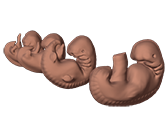
I’m looking at an alien, or at least what looks like one. In reality it’s a human embryo, just a few weeks old; with its strange little legs, a bulging spine, and a pig-like head, it looks more like an insect larva than a human being.
I’m being shown this embryo by John Fredieu, an assistant professor in the department of anatomy. It’s a 3D rendering of a human embryo for a piece of educational software called Embryon, something he has been working on since 2009. It is a project that he hopes will be able to change the way medical students are taught.
Fredieu started the project because he wanted to give professors a better way to teach embryology. As an embryology professor, he has seen daily how difficult it can be for students to learn things from the instructional material currently available. “Right now what they’re using are textbooks, 2D pictures, and sections,” Fredieu told me, pointing to some diagrams. “So they have to reconstruct all the 2D stuff in their head.” When these are the only resources students have, it causes a knowledge gap. “You really have to imagine in 3D to understand what is happening.”
The project was also driven by the medical school instituting their WR2 curriculum in 2006. Under this new program, student learning is focused on clinical experience. While it has been a great success overall, parts of the old system had to be cut. The time that students spend with professors in anatomy, embryology, and histology was slashed down from an average of 170 hours a semester to 60.
This is unacceptable for Fredieu. He is emphatic about how important embryology is.
Pulling up another view of the embryo, he starts pointing at different structures on its face. “All these [pieces] have to come together properly and at the right time in order to create a normal human face. If you have any defect in any one of these parts and how they’re interacting with each other, it can produce: clefts, a loss of tongue, loss of ear, loss of palate; congenital abnormalities. In order for a [doctor] to understand what congenital abnormalities [are present] in a face he is looking at he needs to understand how it’s put together.” This is information that doctors need, but the medical school is not dedicating nearly as much time or resources to actually teach.
To fill that gap, Fredieu wanted to create a way that students could start teaching themselves the material, in addition to what they were getting from professors and textbooks. And so Embryon was born.
Fredieu needed people who could actually complete the project, though, because accurately modeling living things is a technical and time consuming process.
He found the talent he needed when he saw the work of a Cleveland Institute of Art student in one of his classes. “It was after spring break and she came back and brought in a big ball of yarn. And she put this big ball of yarn on the table and started opening it up and it was a placenta with a knitted embryo inside it with a knitted umbilical chord. It was beautiful. And when I saw that I thought, ‘We have these students that are talented like this, why can’t I use them?’”
Fredieu immediately reached out to Amanda Almon, director of biomedical art and animation at CIA and in 2009, with the hard work of two CIA students selected by Almon, the first prototype of what would become Embryon was created.
It was a huge success. Fredieu’s colleagues praised the software and it has been downloaded in 20 different countries.
The software also proved itself as a learning aid. When Fredieu tested it with focus groups, the groups that used the software saw improvements as high as 40 percent over students who had not.
Following that success, Fredieu and Almon decided to take that prototype and make a full featured piece of software, creating something that students at CWRU and other medical schools around the country could use.
Making the team to complete the software was not easy. Artists with both the technical skills and the scientific knowledge necessary to create accurate biological models are very rare. Almon puts it like this: “You don’t always get both in these students. You get ‘I don’t like math,’ or ‘I don’t like science,’ or ‘I’m pretty good at science, but I really like to draw,’ or ‘I’m really awesome at science, but I can’t visualize anything.’ So these students are in this really delicate balance. When we recruit them, it’s a very small class.” How small? By Almon’s estimate, for every class of artists she sees, no more than ten have what it takes to work on this kind of project.
Almon picked out four students for the project: Carly Bartel, Julie Pasini, Jennifer Kerbo, and Maia Garcia Fedor. To help teach the students, Fredieu recruited a former graduate student in anatomy, Story Elliott.
Embryon is more than just a collection of art, though. It is interactive. The user can look at different stages of embryo development. They can pan around and zoom in on different parts of the body, highlight different organs or structures, and see different anomalies and what causes them to develop.
To do all that, they needed someone with programming experience to bring everything together. They turned to Marc Buchner, associate professor in EECS, to advise the project and find a student that could contribute to the programming. He picked out Brendan Mulcahy, a computer science undergrad.
With the team assembled, work started last January.
Now, months later and well into development, Fredieu is optimistic about the project’s future. Feedback has been nothing but positive, and the work that the students are doing is incredible. Within a few weeks of starting, they already had art. Fredieu gestured at the embryo rendering again and said, “I expected some stick figures and they had this.”
The software still has a long way to go though. Months or work and many long nights are still ahead.
Regardless of how long it will take, it’s clear from the feedback and support that Fredieu and his team are receiving that their work is going to make a difference.


