On the bubble
Creatively targeting ovarian tumors
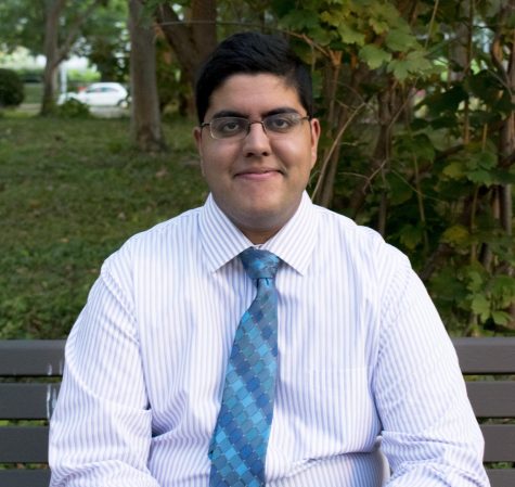
Unlike many students, biomedical engineering major Pavan Kota didn’t have an epiphany senior year of high school or late in college about his future career plans. A junior now, Kota realized his discipline of interest the summer before he joined Case Western Reserve University.
He had been interested in becoming a physician, but was beginning to think that it was the research and devolvement side of medicine that really fascinated him. Not knowing where his first semester would leave him, he had to try to incorporate both. He remembers planning the schedule for his first semester at CWRU, cautious yet fixated.
“There’s something to be said for keeping all your options open,” Kota said. “However, it’s definitely very difficult, and sometimes it can spread you too thin.”
While he did find that he was on the right track, the last thing he expected his first research experience to be with was a children’s plaything: bubbles.
For junior Pavan Kota, these bubbles have been his research focus for the past two years. Kota researches how to attach an antibody to the surface of nano-sized bubbles so that they actively target ovarian cancer cells. The antibody will let the bubble identify a tumor cell, so that the bubbles will target tumors more readily.
These aren’t everyday bubbles, however. The nanometer size and the fluorocarbon gases they contain make them unique. They reflect sound waves back at their sources, a useful trait in the medical world. When these bubbles are injected into the bloodstream, they end up congregating in tumors. Then, an ultrasound can be used to identity the tumor. If there is a chemotherapy drug in the bubble, the ultrasound can actually be used to pop it and deliver the drug.
Currently microscale bubbles are used to indirectly identify or treat tumors. Kota explained that bubbles accumulate more in tumor cells, since their cell membranes contain larger pores. Current diagnosis and treatment ultrasounds rely on this fact.
However, many of these pores are actually thousands of times smaller, and cancer cells can have high pressures that may interfere with bubbles entering and remaining inside them. The lab is trying to develop a smaller and active-targeting bubble to provide better ultrasound pictures.
The lab’s work has primarily been attaching the antibody to the bubble and confirming its presence. For Kota, that meant a lot of training at first.
“There’s a lot to be said for getting involved in research as early as possible,” he said.
In each experiment, Kota attempted to attach the antibody to the bubble in a certain set of chemical conditions. He then had to look at whether he attached the antibody to the bubble successfully.
To do this, Kota used a second antibody, which has a fluorescent molecule attached to it. It can attach to the first antibody, which is attached to the bubble at the same time. He simply adds the second to the first and does a wash. If the second attaches to the first, the second remains and Kota can confirm this through the fluorescent signal.
Kota also demonstrated that the antibody-bubble complexes bind to tumor cells that are grown in his lab on petri dishes.
This semester, Kota is on a co-op working at General Electric. He isn’t sure if industry or academia is the right place for him. While there is more time now, when compared to that first summer when he chose his first-year classes, this decision is perhaps more critical. It waits for him in the near future.
“Getting hands-on experience can help guide your decisions and make it happen as quickly as possible,” Kota said.

Kushagra Gupta is a cognitive science and biology student and is working towards a masters in medical physiology. He's served as The Observer’s The Director...

